X-ray of rheumatoid arthritis in the feet
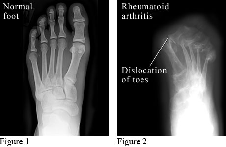
Figure 1 courtesy of Intermountain Medical Imaging, Boise, Idaho. Figure 2 courtesy of Paul Traughber, MD, Boise, Idaho.
The X-ray on the left shows a normal foot.
The X-ray on the right shows a foot with advanced rheumatoid arthritis. Cartilage and bone are worn away, and the bones of the toes have moved out of their normal positions.
Current as of: July 10, 2023
Author: Healthwise Staff
Clinical Review Board
All Healthwise education is reviewed by a team that includes physicians, nurses, advanced practitioners, registered dieticians, and other healthcare professionals.